Penile curvature, whether congenital or related to Peyronie’s disease, can cause significant functional impairment and impair sexual quality of life. When the curvature stabilizes and medical treatment is no longer indicated, surgery becomes the standard solution for restoring functional straightness to the penis.
As urological and andrological surgeons, the goal is to adapt the surgical technique to each patient’s anatomy and the characteristics of their curvature. Here are the two main surgical approaches we use: tunica albuginea plication (Nesbit technique) and incision with patch placement (TachoSil).
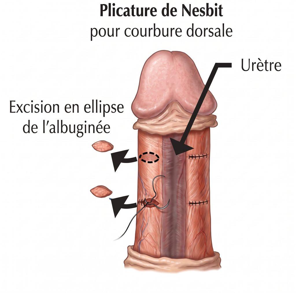
1. The Plicature of the Tunica albuginea (Nesbit’s Technique)
This approach is based on a simple geometric principle: to straighten the penis, the longer side (the convex side) is shortened to align it with the shorter side (the concave side, where the fibrous plaque is usually located in the case of Peyronie’s disease).
During the procedure, small elliptical incisions or excisions are made in the tunica albuginea (the sheath surrounding the corpora cavernosa) on the side opposite the curvature. The edges are then sutured with thread, which straightens the penis.
- Indications: Moderate curvatures (generally less than 60 degrees), absence of hourglass deformity, and patients with an adequate initial penis length.
- Advantages: Controlled surgical technique, shorter operating time, preservation of erectile function (very low risk of postoperative erectile dysfunction).
- Disadvantages: Causes an inevitable shortening of the penis, proportional to the degree of the initial curvature.
2. Incision with Graft (TachoSil Patch)
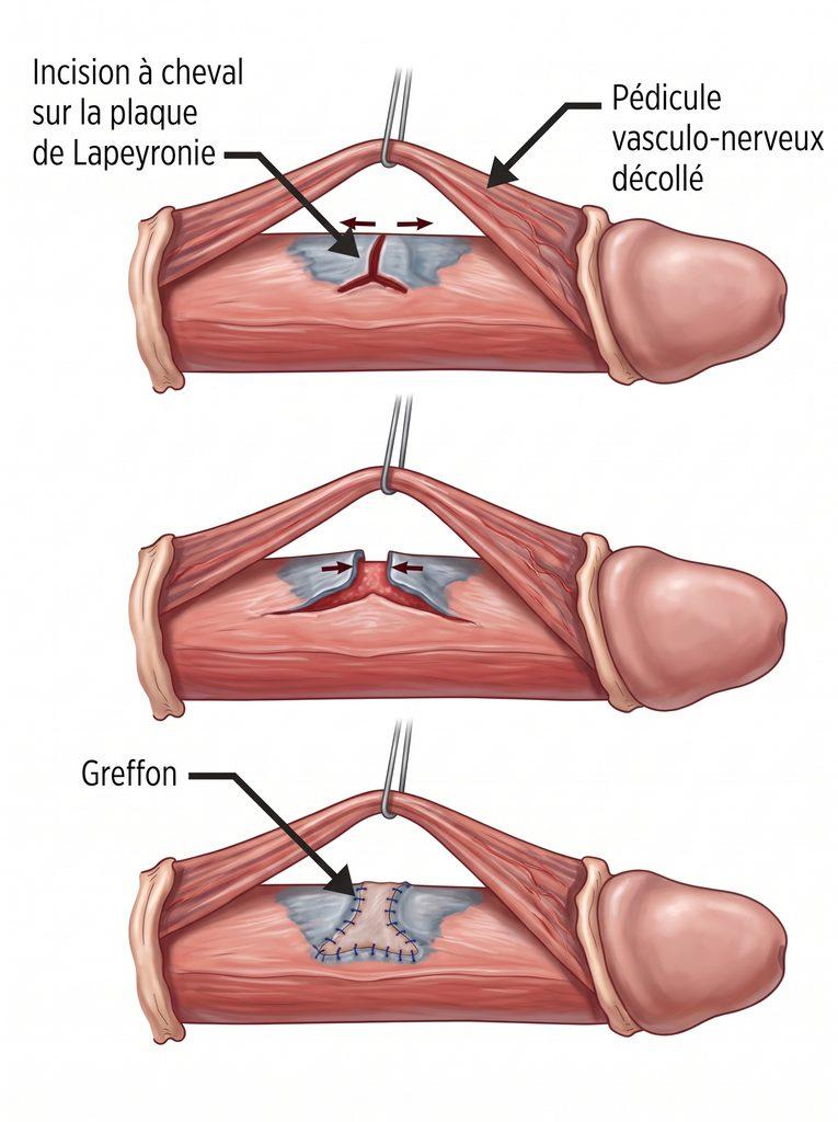
Unlike plication, this approach aims to lengthen the shorter side. The principle is to directly section the fibrotic plaque in the case of Peyronie’s disease, or the tunica albuginea on the concave side in the case of congenital curvature.
This incision creates an opening (a defect) in the tunica albuginea, which must be filled. For this, we use a covering patch. TachoSil , an equine collagen matrix coated with human coagulation factors (fibrinogen and thrombin), is currently a material of choice. It adheres strongly to tissues, ensures excellent sealing, and promotes tissue healing without requiring complex sutures.
- Indications: Severe curvatures (greater than 60 degrees), complex deformities (hourglass, hinge), and patients with a short penis where further shortening would be detrimental.
- Advantages: Allows for the straightening of even the most complex curvatures while minimizing the loss of penis length.
- Disadvantages: More complex surgical procedure, often requiring dissection of the neurovascular bundle. Higher risk of temporary alteration of glans sensitivity and de novo erectile dysfunction (sometimes requiring postoperative pharmacological support). Tissue rehabilitation with traction for several months is essential postoperatively.
CLINICAL CASES:
Clinical Case 1: Treatment of congenital penile curvature by Nesbit plication
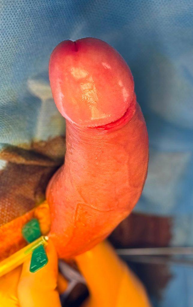
Background and Presentation: A 16-year-old patient, accompanied by his parents, was seen for consultation regarding a penile deformity that had become evident with pubertal development. The clinical examination at rest was entirely normal. Analysis of photographs taken by the patient during erection (self-photographs essential for diagnosis) revealed a significant left lateral curvature.
Unlike Peyronie’s disease, this deformity is not associated with pain or palpable fibrous plaques. It is a congenital curvature due to an asymmetrical development of the tunica albuginea, the membrane surrounding the corpora cavernosa: the right side develops longer than the left. While erectile function is otherwise perfect, the psychological impact is significant, and the angle could make future sexual intercourse complicated, even impossible or painful for the partner.
Surgical Rationale
In cases of congenital curvature in patients with satisfactory penile length, the standard technique is shortening the convex side (the longer side). The goal is to symmetrize the corpora cavernosa. The Nesbit technique is preferred here for its geometric reliability, long-term stability, and excellent rate of preserving erectile function.
Procedure for the Intervention (Nesbit Technique)
The procedure is performed on an outpatient basis (same-day discharge), under general anesthesia, and follows several standardized steps:
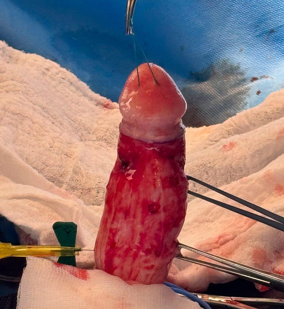
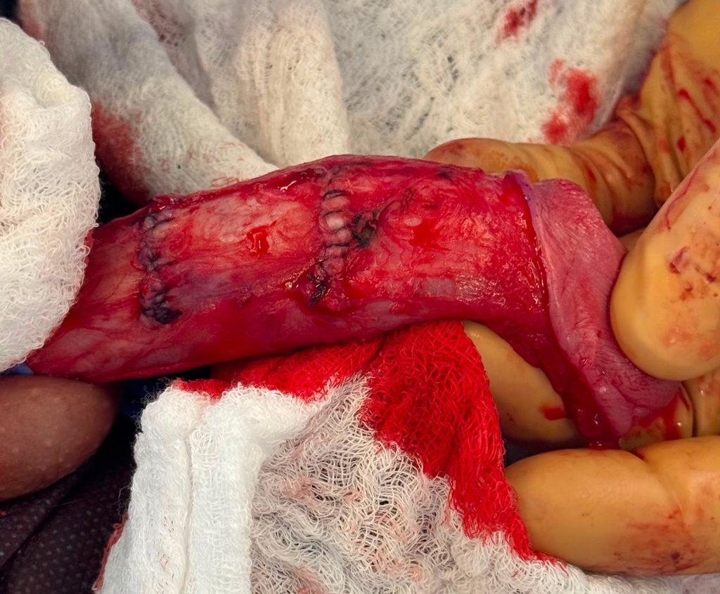
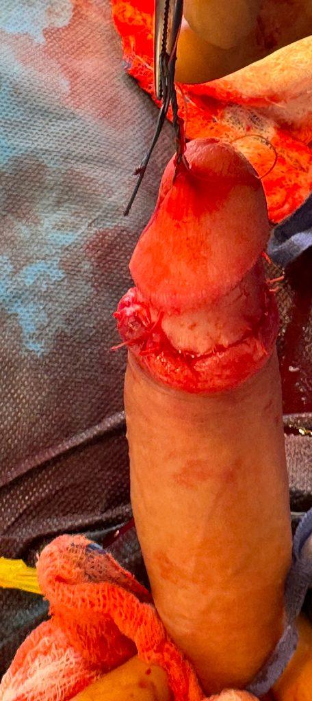
- Anatomical exposure: A circular incision under the glans allows the skin of the penis to be "ungloved" up to its base in order to expose the tunica albuginea of the corpora cavernosa over its entire circumference.
- Artificial erection: Injecting saline solution directly into the corpora cavernosa allows a rigid erection to be recreated on the operating table.
- Neurovascular dissection: To work on the tunica albuginea safely, the dorsal neurovascular band (which provides sensitivity to the glans) is carefully dissected and temporarily removed.
- Excision and Suturing (Nesbit Technique): Initially, locating clips are placed on the tunica albuginea. Once the fold areas are marked, small ellipses of tunica albuginea (approximately 5 to 10 mm wide) are surgically removed from the longer side (in this case, the right). The edges of these incisions are then brought together and closed with absorbable sutures.
- Check-up: A second artificial erection is induced to confirm the penis is perfectly straight. If the result is perfect, the skin is repositioned and sutured with absorbable thread.
Post-operative effects and long-term outcome
The immediate postoperative period is marked by typical swelling. Involuntary nocturnal erections may be painful for the first few days. The main instruction is strict sexual abstinence (including masturbation) for 6 weeks to ensure the sutures heal properly.
Clinical Case 2: Treatment of a severe congenital ventral curvature (90°) by incision and TachoSil patch
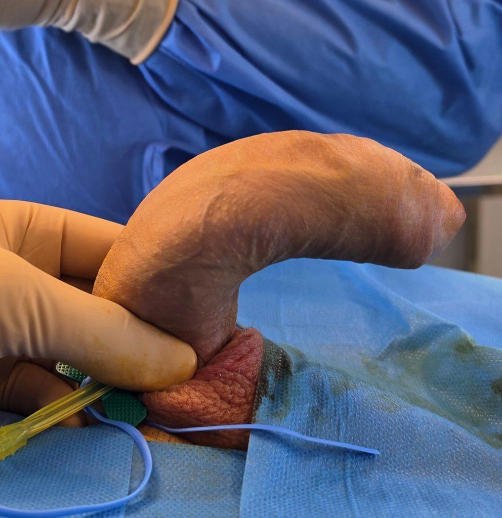
Clinical Context: We are seeing a 21-year-old man in consultation with a congenital ventral curvature of the penis. The patient describes a very pronounced downward angulation, which appeared at puberty and makes any attempt at penetration physically impossible and psychologically distressing. The clinical examination in the flaccid state (at rest) is normal; the urethra is well positioned at the tip of the glans (absence of hypospadias). Evaluation based on photographs of the erect penis reveals a ventral curvature estimated at nearly 90 degrees. Erectile function is otherwise excellent.
Surgical Rationale: Why rule out plication?
In cases of congenital curvature, the plication technique (shortening the longer side, in this case the dorsal side) is often preferred for its simplicity. However, the greater the angle of curvature, the greater the loss of length resulting from the plication will be.
For a curvature reaching 90 degrees, a dorsal plication would result in an anatomically and functionally unacceptable penile shortening. The surgical strategy therefore focuses on lengthening the shorter side (the ventral side). This approach requires incising the ventral tunica albuginea to straighten the penis, then filling the resulting space with a graft.
The complexity of this approach lies in the anatomy: the area to be incised is covered by the corpus spongiosum, the structure that contains the urethra.
Procedure of the Intervention
The procedure is performed under general anesthesia. To guarantee a perfect anatomical result and preserve vital structures, the procedure follows rigorous steps:
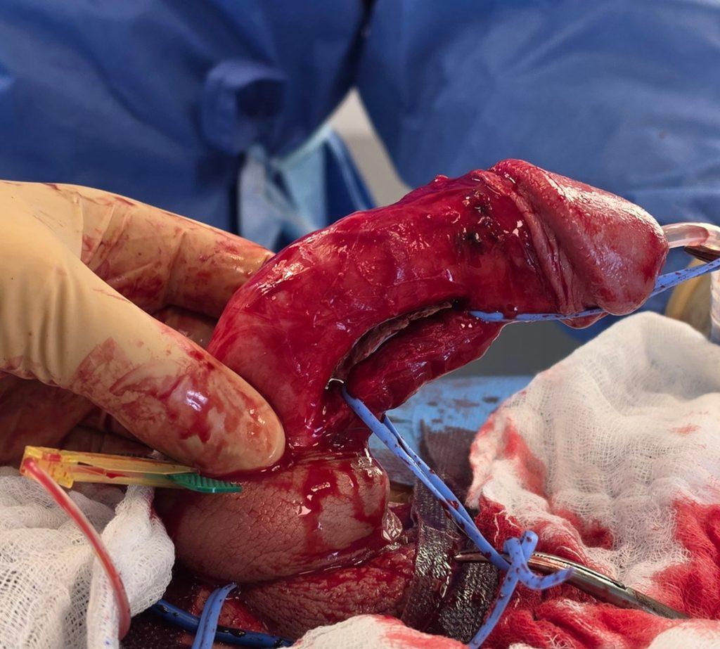
- Surgical approach and localization : A circumferential incision is made under the glans, and the gloves are removed to expose the corpora cavernosa. An artificial erection (injection of saline solution into the cavernosa) is performed to confirm the 90-degree angle and precisely mark the point of maximum curvature on the ventral surface.
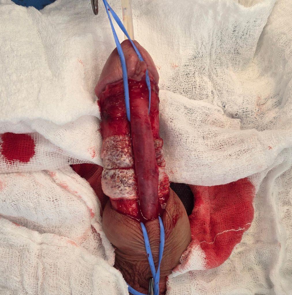
- Urethral release: The corpus spongiosum (containing the urethra) is closely attached to the ventral surface of the corpora cavernosa. It is dissected and mobilized to detach it from the tunica albuginea.
- Tunica albuginea incision: A transverse incision is made on the ventral tunica albuginea, at the apex of the curvature. This tissue release allows for immediate straightening of the penile axis. The separation of the edges creates a defect that must now be filled.
- Graft placement (TachoSil): The defect is measured. A TachoSil patch (equine collagen matrix coated with coagulation factors) is custom-cut. Applied to the tunica albuginea defect, it adheres firmly to the tissues due to moisture and ensures a perfect seal of the corpora cavernosa without requiring multiple sutures that could compromise tissue elasticity.
- Verification and closure: The urethra is repositioned in its natural anatomical bed, and the skin layers are sutured.
Post-operative care and rehabilitation protocol:
- A total sexual abstinence period of 6 weeks is prescribed. This period is essential to allow the TachoSil patch to integrate before being completely absorbed, leaving behind a supple and strong new tunica albuginea.
- Mechanical rehabilitation: the patient begins a penile traction rehabilitation protocol (extensor or vacuum) as soon as possible for 6 months . This mechanical therapy is essential after incision-graft surgery: it guides tissue healing, prevents scar retraction of the graft, and maximizes the preservation of penile length.
Clinical Case 3: Treatment of severe spinal curvature (Peyronie’s disease) by incision and TachoSil patch
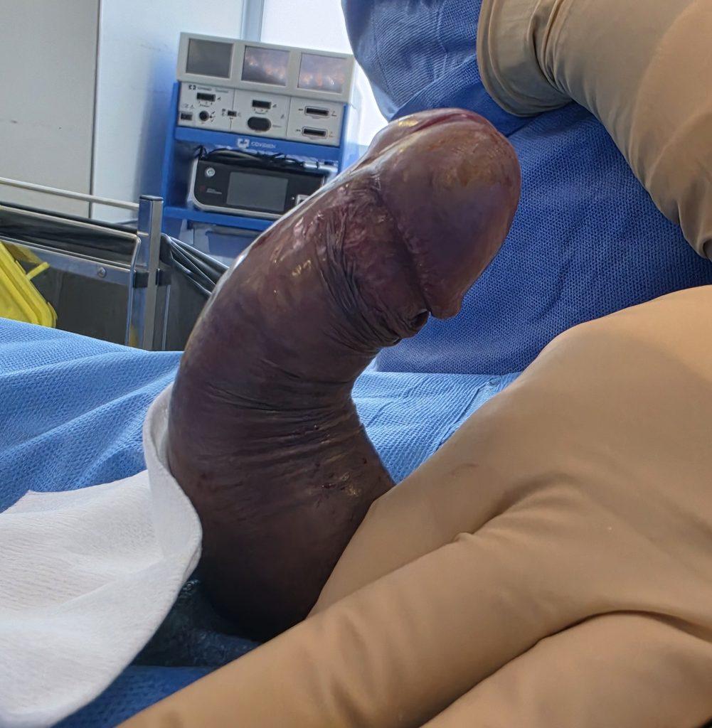
Clinical Context: We are seeing a 52-year-old patient presenting with an acquired penile deformity that has been developing for 18 months. This is Peyronie’s disease, now in the sequelae phase (the deformity has been stable for over 6 months and pain during erections has disappeared). Clinical examination reveals a palpable fibrous plaque on the dorsal aspect of the penis. Photographs taken during erection show a severe dorsal (upward) curvature, measuring 70 degrees. This angulation makes penetration very difficult and uncomfortable for both partners. The key point of the preoperative evaluation is the preservation of excellent erectile function, which allows us to direct the patient towards surgery for pure correction, without the need for a penile prosthesis.
Surgical Rationale: The Choice of Incision-Graft
Faced with a 70-degree dorsal curvature, the plication technique (shortening the healthy ventral surface) would correct the axis, but at the cost of a significant loss of length. The objective here is therefore to restore the length of the dorsal surface, which is affected by the disease.
This approach requires incising the fibrous plaque. The anatomical challenge lies in the location of this plaque: it is located under the dorsal neurovascular band , the complex structure that runs along the back of the penis and provides vascularization and sensory innervation to the glans.
Procedure of the Intervention
Performed under general anesthesia, this procedure uses micro-dissection techniques to preserve nerve structures:
- Exposure and evaluation: A circumferential approach allows the penis to be "ungloved". An artificial erection with saline solution precisely determines the 70-degree angle and the location of maximum retraction.
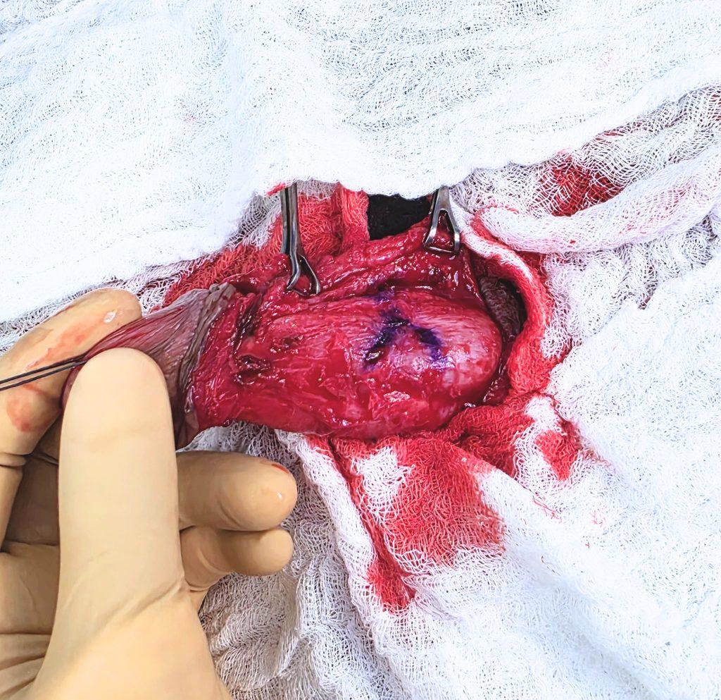
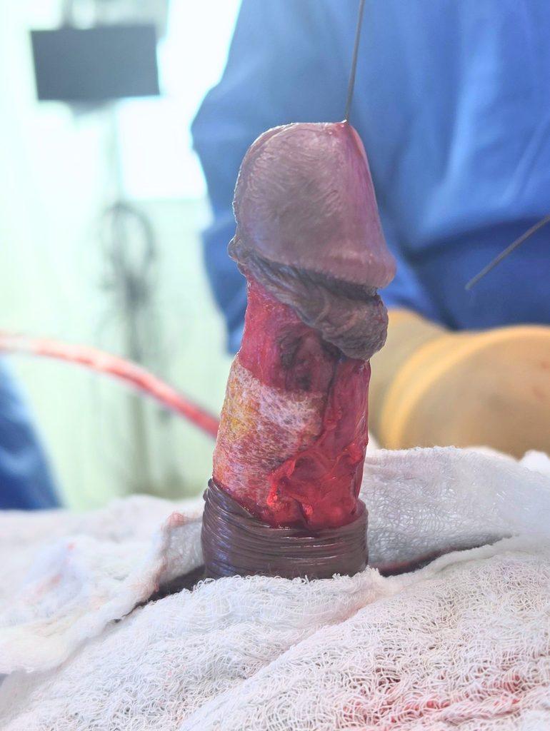
- Dissection of the neurovascular bundle: Using magnifying loupes, the dorsal neurovascular bundle is carefully separated from the underlying tunica albuginea and lifted laterally. This maneuver allows safe exposure of the fibrous plaque.
- Plaque incision: A transverse incision in the shape of two inverted Ys is made directly into the fibrous tissue. Release is immediate and the axis of the penis straightens, revealing an albuginea-cavernosus defect (an opening) at the incision site.
- Closure with a graft (TachoSil): The defect is filled by applying a TachoSil patch (collagen matrix coated with coagulation factors). This biomaterial, which is completely absorbable , adheres physiologically to the tissues and ensures the seal of the corpora cavernosa without requiring sutures (which could damage the tissues and elasticity).
- The neurovascular strip is delicately repositioned in its natural axis over the graft, and the skin layers are closed.
Post-Operative Care and Rehabilitation Protocol
The procedure is performed on an outpatient basis or requires a short overnight stay. Preserving the length and quality of the final result depend on a rigorous post-operative protocol:
- Healing phase: Total sexual abstinence for 6 weeks is imperative. This is the time required for the TachoSil patch to integrate with the cavernous tissues before its complete absorption, and for the dissected micro-vessels and nerves to recover.
- Tissue rehabilitation phase: As soon as possible, the patient begins mechanical rehabilitation using a penile extender or vacuum device for 6 months . This step is crucial after a graft: it helps guide healing, prevent scar retraction of the new tissue, and optimize length gain.
In conclusion, as we have seen, every patient is different. To guarantee the best possible result, it is essential to master all surgical techniques in order to adapt to each situation and offer a personalized treatment plan. This is why it is important to consult a surgeon specializing in the penis before considering any procedure.